Figure 8
400 × 348px
· 46,0 Kio
Source Paper
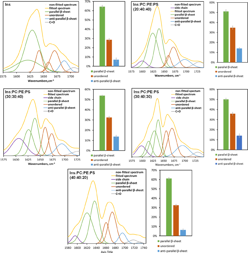
Concentration of Phosphatidylserine Influence Rates of Insulin Aggregation and Toxicity of Amyloid Aggregates In Vitro.Cite This Figure
 > Source: Mikhail Matveyenka et al. "Concentration of Phosphatidylserine Influence Rates of Insulin Aggregation and T." *ACS chemical neuroscience*, 2023. PMID: [37279439](https://pubmed.ncbi.nlm.nih.gov/37279439/)
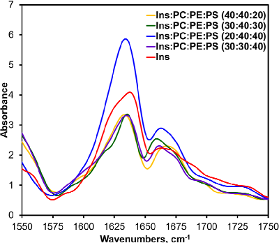
<figure>
  <img src="https://pdfs.citedhealth.com/figures/37279439/150.png" alt="Figure 4. AFM-IR spectra acquired from insulin (Ins) fibrils grown in the lipid-free environment (red), insulin in the presence of LUVs of PC/PE/PS (40:40:20) (yellow), PC/PE/PS (30:40:30) (green), PC/ PE/PS (20:40:40) (blue), and PC/PE/PS (30:30:40) (pur" />
  <figcaption>Figure 8. Figure 4. AFM-IR spectra acquired from insulin (Ins) fibrils grown in the lipid-free environment (red), insulin in the presence of LUVs of PC/PE/PS (40:40:20) (yellow), PC/PE/PS (30:40:30) (green), PC/ PE/PS (20:40:40) (blue), and PC/PE/PS (30:30:40) (pur<br>  Source: Mikhail Matveyenka et al. "Concentration of Phosphatidylserine Influence Rates of Insulin Aggregation and T." <em>ACS chemical neuroscience</em>, 2023. PMID: <a href="https://pubmed.ncbi.nlm.nih.gov/37279439/">37279439</a></figcaption>
</figure>