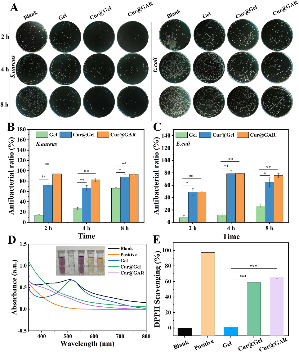
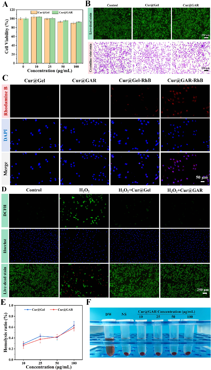
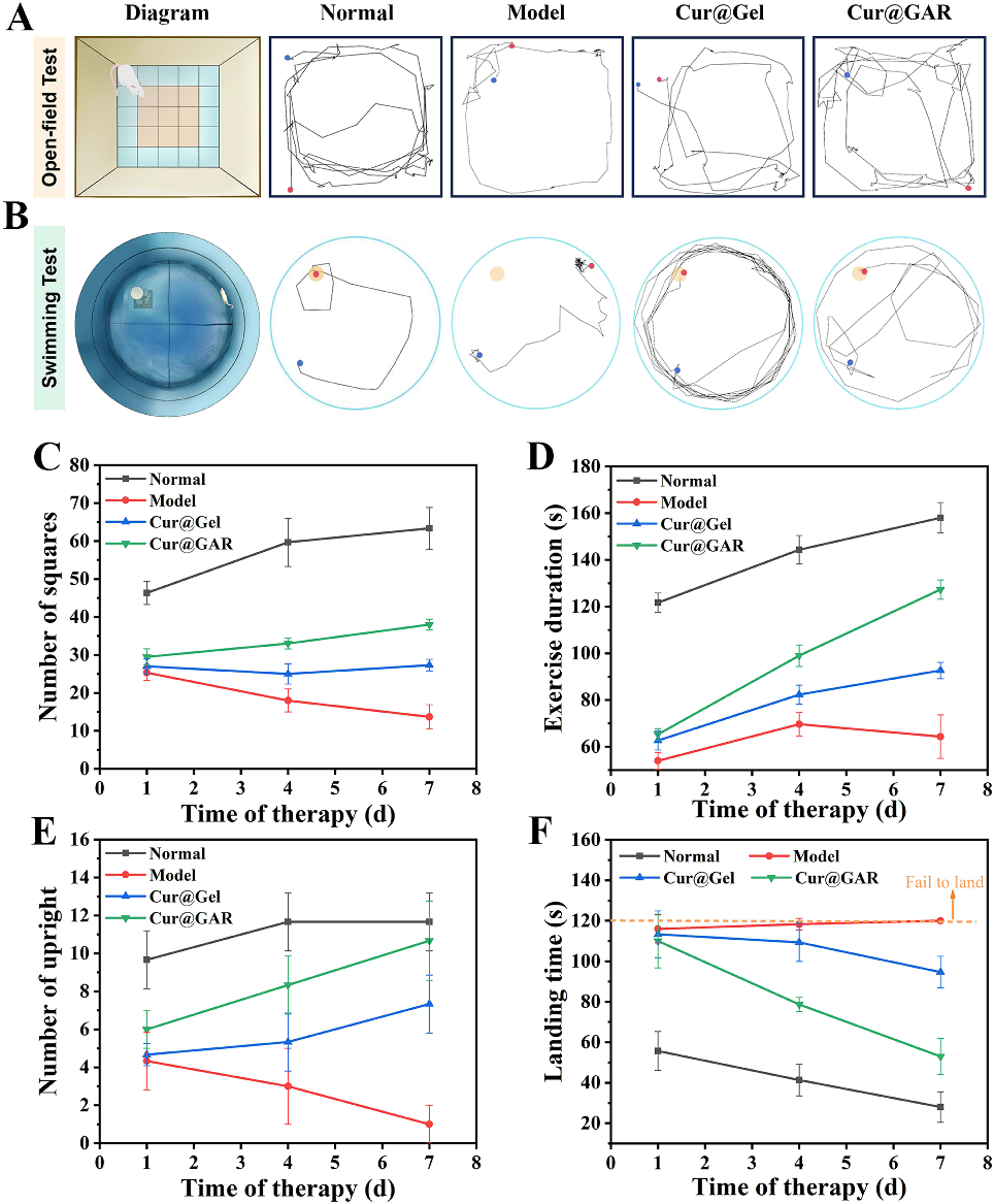
Figure 7
720 × 825px
· 657,4 Kio
Source Paper
Curcumin-Loaded Gelatin Nanoparticles Cross the Blood-Brain Barrier to Treat Ischemic Stroke by Attenuating Oxidative Stress and Neuroinflammation.Cite This Figure
 > Source: Qinglu Yang et al. "Curcumin-Loaded Gelatin Nanoparticles Cross the Blood-Brain Barrier to Treat Isc." *International journal of nanomedicine*, 2024. PMID: [39553455](https://pubmed.ncbi.nlm.nih.gov/39553455/)
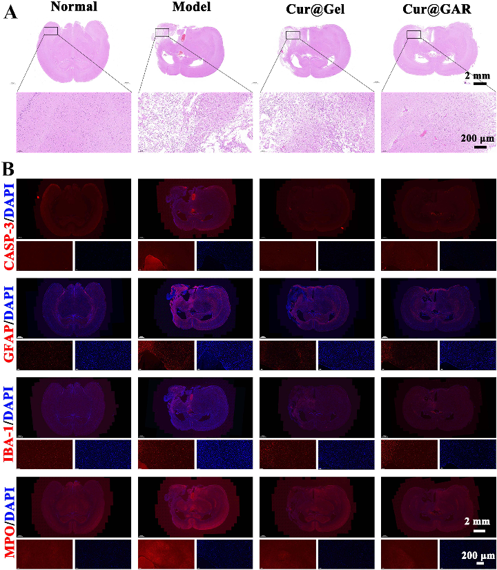
<figure> <img src="https://pdfs.citedhealth.com/figures/39553455/233.png" alt="Figure 5 (A) H&E staining images of brain tissue sections from experimental rats on day 7 after surgery. (B) Immunofluorescence staining images of CASP-3, GFAP, IBA-1, and MPO in brain tissue sections of experimental rats on day 7 after surgery." /> <figcaption>Figure 7. Figure 5 (A) H&E staining images of brain tissue sections from experimental rats on day 7 after surgery. (B) Immunofluorescence staining images of CASP-3, GFAP, IBA-1, and MPO in brain tissue sections of experimental rats on day 7 after surgery.<br> Source: Qinglu Yang et al. "Curcumin-Loaded Gelatin Nanoparticles Cross the Blood-Brain Barrier to Treat Isc." <em>International journal of nanomedicine</em>, 2024. PMID: <a href="https://pubmed.ncbi.nlm.nih.gov/39553455/">39553455</a></figcaption> </figure>