Alpha-Lipoic Acid Рисунки
10 иллюстрации из рецензируемых исследований
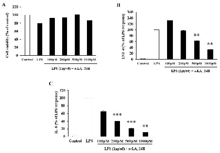
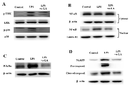
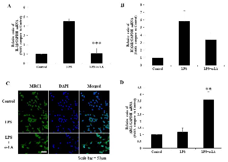
Effects of α-lipoic acid on LPS-induced neuroinflammation and NLRP3 inflammasome activation through …
Effects of α-lipoic acid on LPS-induced neuroinflammation and NLRP3 inflammasome activation through …
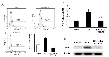
Effects of α-lipoic acid on LPS-induced neuroinflammation and NLRP3 inflammasome activation through …
Effects of α-lipoic acid on LPS-induced neuroinflammation and NLRP3 inflammasome activation through …
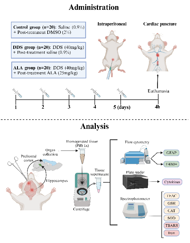
Behavioral or neurological assessment data from a study evaluating alpha-lipoic acid's neuroprotective effects against dapsone-induced neuroinflammation and oxidative stress in an animal model.
Alpha-Lipoic Acid Reduces Neuroinflammation and Oxidative Stress Induced by Dapsone in an …
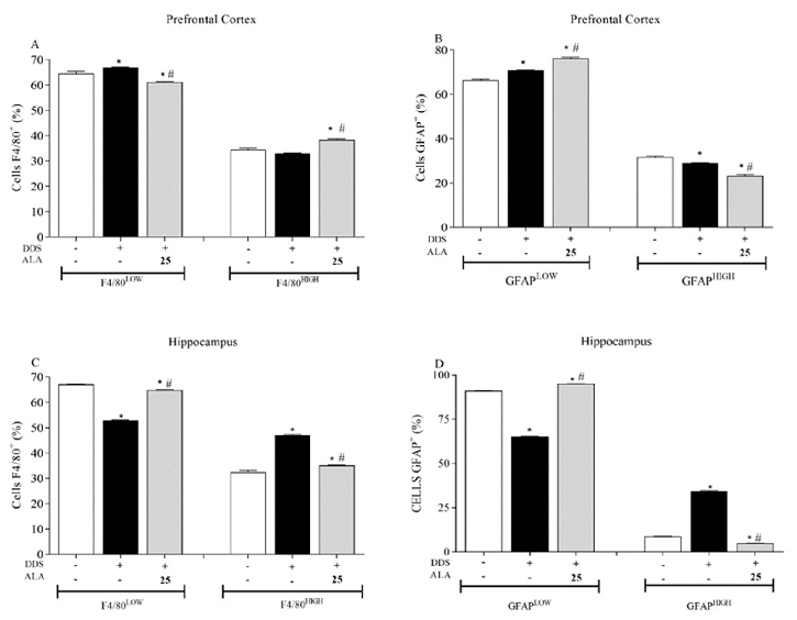
Brain tissue analysis showing markers of neuroinflammation in dapsone-treated animals, comparing alpha-lipoic acid-supplemented versus control groups.
Alpha-Lipoic Acid Reduces Neuroinflammation and Oxidative Stress Induced by Dapsone in an …
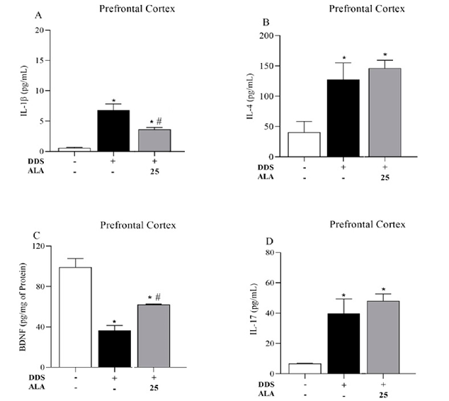
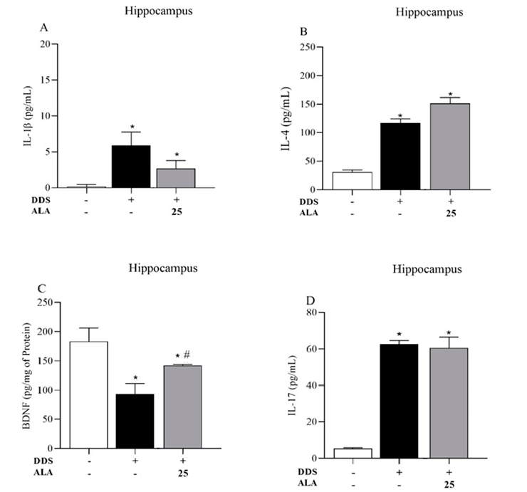
Cytokine levels (IL-1beta, IL-6, and related markers) in prefrontal cortex tissue of dapsone-treated animals, demonstrating alpha-lipoic acid's anti-inflammatory effects.
Alpha-Lipoic Acid Reduces Neuroinflammation and Oxidative Stress Induced by Dapsone in an …
Histological or immunohistochemical analysis of brain tissue from the alpha-lipoic acid and dapsone neuroinflammation study.
Alpha-Lipoic Acid Reduces Neuroinflammation and Oxidative Stress Induced by Dapsone in an …
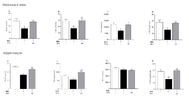
Antioxidant capacity measurements (TEAC, GSH, SOD, and CAT) in prefrontal cortex tissue, showing alpha-lipoic acid's ability to counteract dapsone-induced oxidative stress.
Alpha-Lipoic Acid Reduces Neuroinflammation and Oxidative Stress Induced by Dapsone in an …
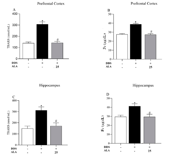
Thiobarbituric acid reactive substances (TBARS) measurements indicating lipid peroxidation levels in dapsone-treated animals, with and without alpha-lipoic acid intervention.
Alpha-Lipoic Acid Reduces Neuroinflammation and Oxidative Stress Induced by Dapsone in an …