Description
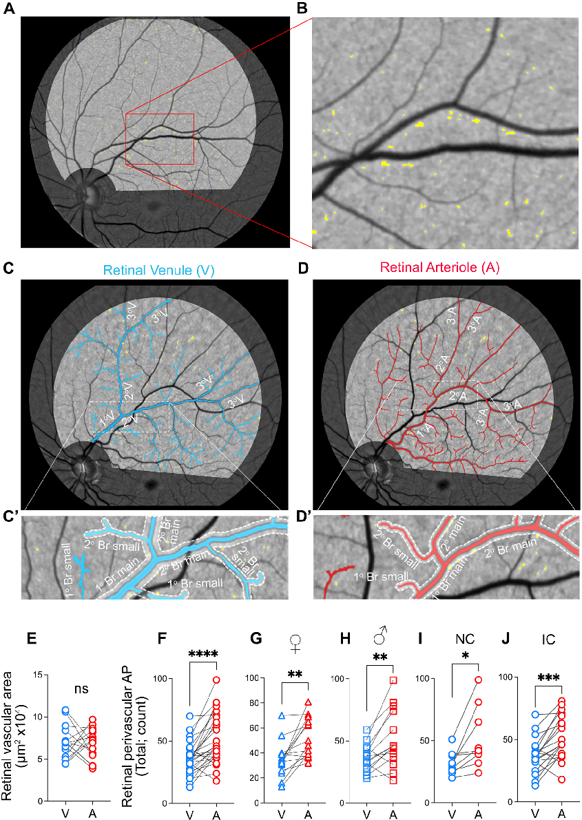
Measured parameters from a study evaluating retinal peri, contributing to the overall assessment of the relationship between amyloidosis and vasculature in cognitive impairment and Alzheimer's disease (AD) pathogenesi.
More Figures from This Paper
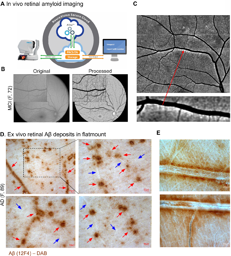
Figure 2
Statistical analysis from research investigating retinal peri, comparing treatment groups and control conditions.
chart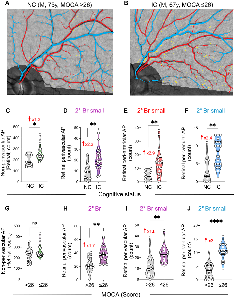
Figure 4
Graphical representation of outcomes in a study of retinal peri, highlighting trends observed across experimental conditions.
chart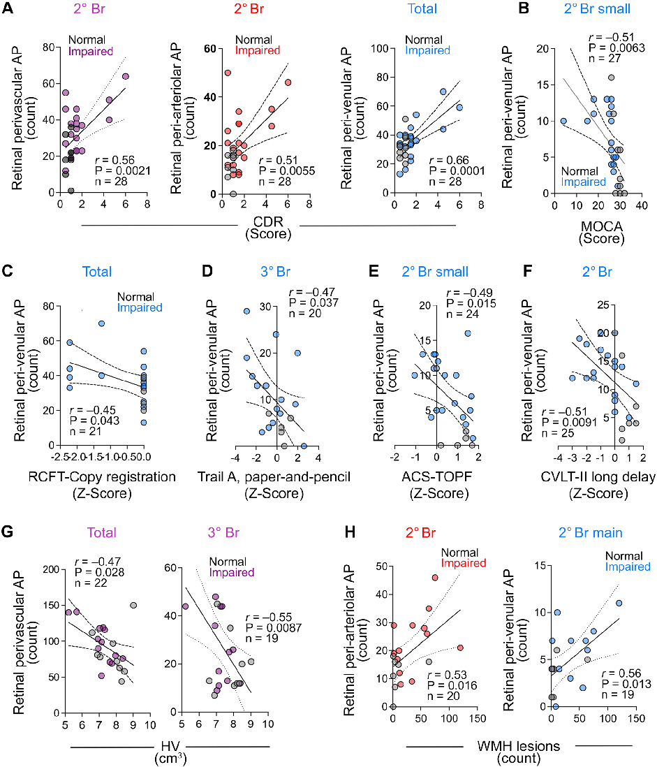
Figure 5
Fig. 4 Correlations between retinal perivascular amyloid plaque distribution with cognitive and neuroimaging measures.
chartFigure 3
Chart
845 × 1191px
· 766.1 KB
Source Paper
Retinal peri-arteriolar versus peri-venular amyloidosis, hippocampal atrophy, and cognitive impairment: exploratory trial.Cite This Figure
 > Source: Oana M Dumitrascu et al. "Retinal peri-arteriolar versus peri-venular amyloidosis, hippocampal atrophy, an." *Acta neuropathologica communications*, 2024. PMID: [38943220](https://pubmed.ncbi.nlm.nih.gov/38943220/)
<figure>
  <img src="https://pdfs.citedhealth.com/figures/38943220/83.png" alt="Measured parameters from a study evaluating retinal peri, contributing to the overall assessment of the relationship between amyloidosis and vasculature in cognitive impairment and Alzheimer's disease (AD) pathogenesi." />
  <figcaption>Figure 3. Measured parameters from a study evaluating retinal peri, contributing to the overall assessment of the relationship between amyloidosis and vasculature in cognitive impairment and Alzheimer's disease (AD) pathogenesi.<br>  Source: Oana M Dumitrascu et al. "Retinal peri-arteriolar versus peri-venular amyloidosis, hippocampal atrophy, an." <em>Acta neuropathologica communications</em>, 2024. PMID: <a href="https://pubmed.ncbi.nlm.nih.gov/38943220/">38943220</a></figcaption>
</figure>