Curcumin الأشكال
9 أشكال من أبحاث محكّمة
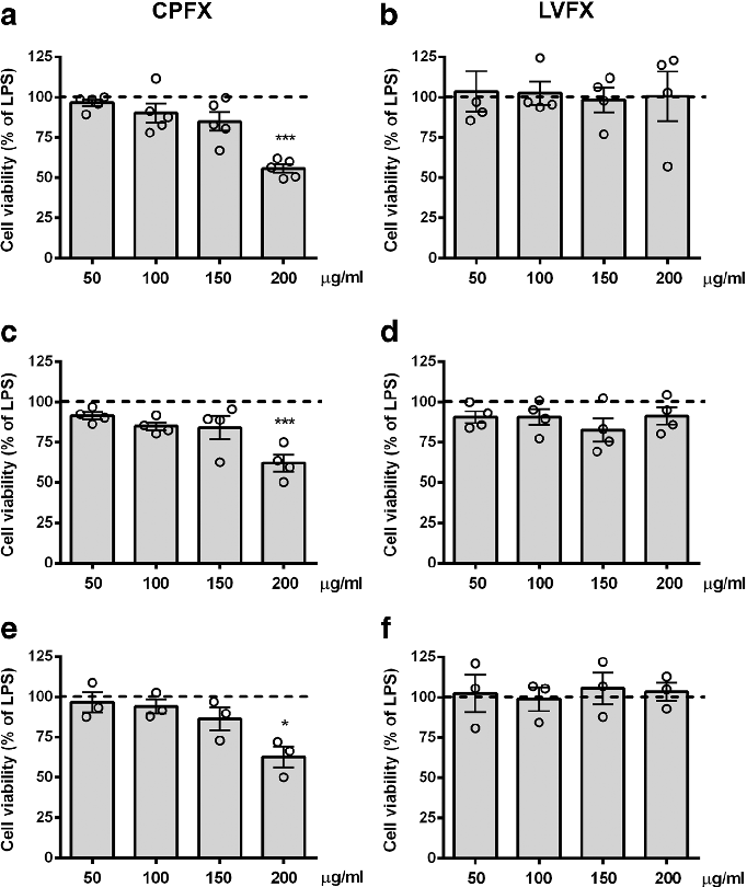
Cell viability assays demonstrate that ciprofloxacin and levofloxacin at the tested concentrations do not significantly reduce microglial survival, confirming that anti-inflammatory effects are not due to cytotoxicity.
Ciprofloxacin and levofloxacin attenuate microglia inflammatory response via TLR4/NF-kB pathway.
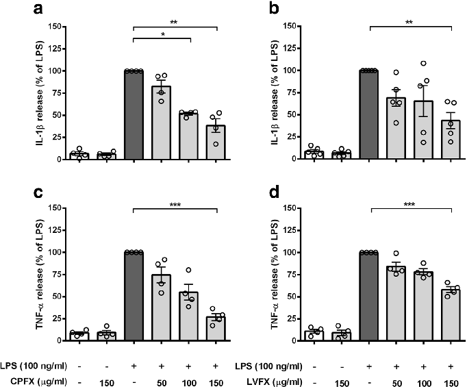
Cytokine release profiles from LPS-stimulated cortical microglia reveal dose-dependent reductions in TNF-alpha and IL-6 following fluoroquinolone treatment.
Ciprofloxacin and levofloxacin attenuate microglia inflammatory response via TLR4/NF-kB pathway.
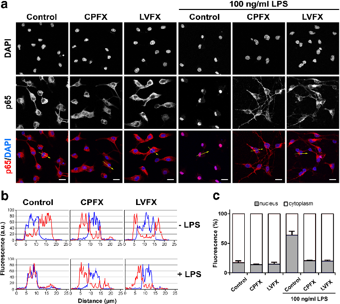
NF-kB nuclear translocation in LPS-stimulated microglia is attenuated by both ciprofloxacin and levofloxacin, as shown by immunofluorescence or reporter gene assays.
Ciprofloxacin and levofloxacin attenuate microglia inflammatory response via TLR4/NF-kB pathway.
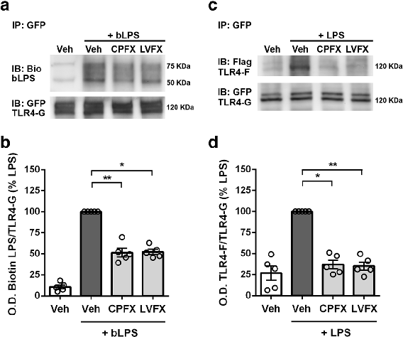
LPS binding and TLR4 dimerization assays in Ba/F3 cells demonstrate that fluoroquinolones interfere with the initial receptor activation step of innate immune signaling.
Ciprofloxacin and levofloxacin attenuate microglia inflammatory response via TLR4/NF-kB pathway.
Statistical analysis from research investigating retinal peri, comparing treatment groups and control conditions.
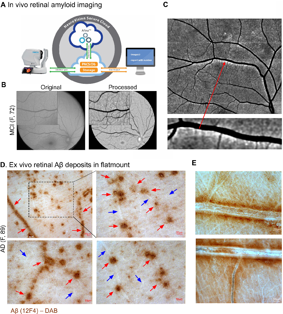
Retinal peri-arteriolar versus peri-venular amyloidosis, hippocampal atrophy, and cognitive impairment: exploratory trial.
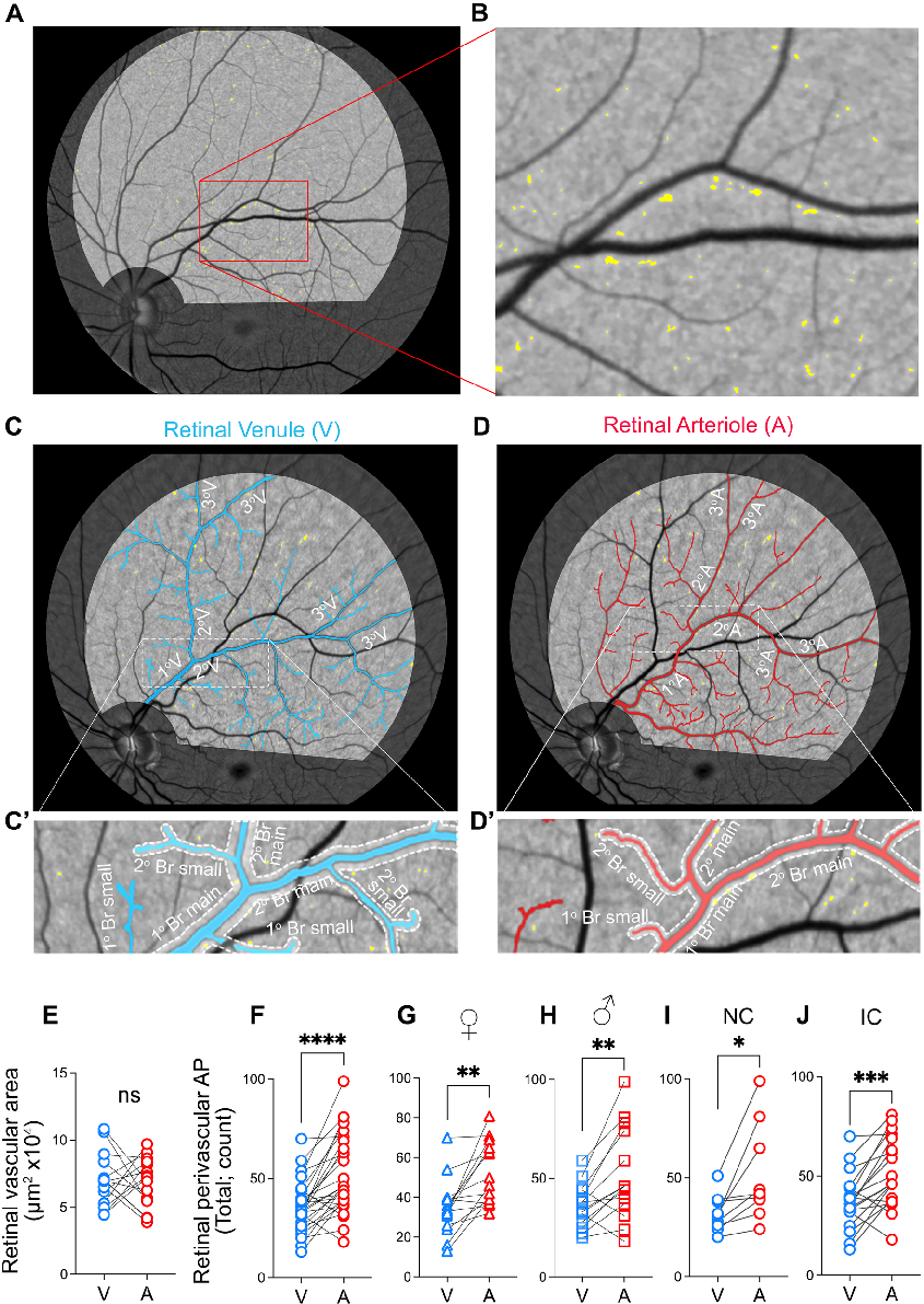
Measured parameters from a study evaluating retinal peri, contributing to the overall assessment of the relationship between amyloidosis and vasculature in cognitive impairment and Alzheimer's disease (AD) pathogenesi.
Retinal peri-arteriolar versus peri-venular amyloidosis, hippocampal atrophy, and cognitive impairment: exploratory trial.
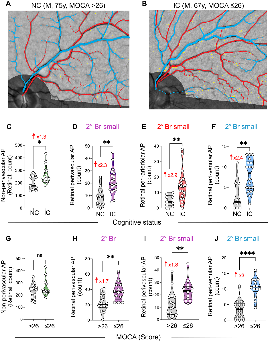
Graphical representation of outcomes in a study of retinal peri, highlighting trends observed across experimental conditions.
Retinal peri-arteriolar versus peri-venular amyloidosis, hippocampal atrophy, and cognitive impairment: exploratory trial.
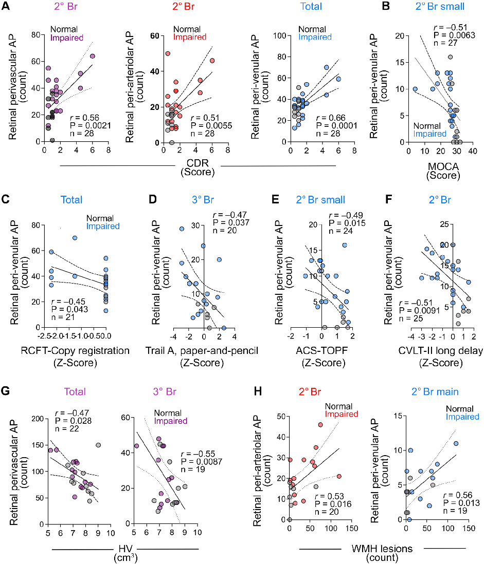
Fig. 4 Correlations between retinal perivascular amyloid plaque distribution with cognitive and neuroimaging measures.
Retinal peri-arteriolar versus peri-venular amyloidosis, hippocampal atrophy, and cognitive impairment: exploratory trial.
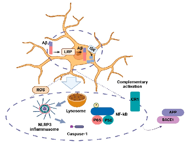
Brain microglia activation in AD. Microglia are effective in Aβ clearance, neuroinflammation, and the production and aggregation of Aβ.
Mechanisms Linking Obesity, Insulin Resistance, and Alzheimer's Disease: Effects of Polyphenols and …