विवरण
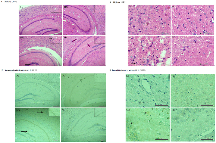
Immunofluorescence staining of amyloid-beta plaques in the temporal cortex and hippocampus shows reduced plaque burden in nutrient-treated APP-PSN transgenic mice.
More Figures from This Paper
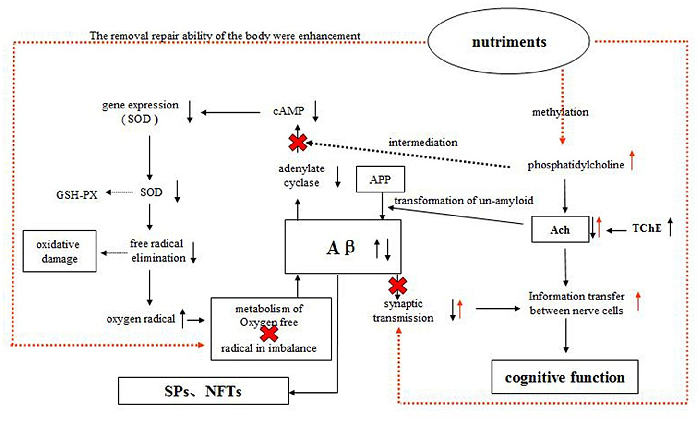
Figure 4
A schematic diagram illustrates the proposed mechanisms by which the compound nutrient mixture may protect against Alzheimer's pathology, including acetylcholine and cAMP signaling.
diagram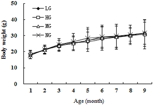
Figure 5
Daily food intake across groups confirms consistent consumption, with no significant differences between the low, high, and model groups during the study.
chart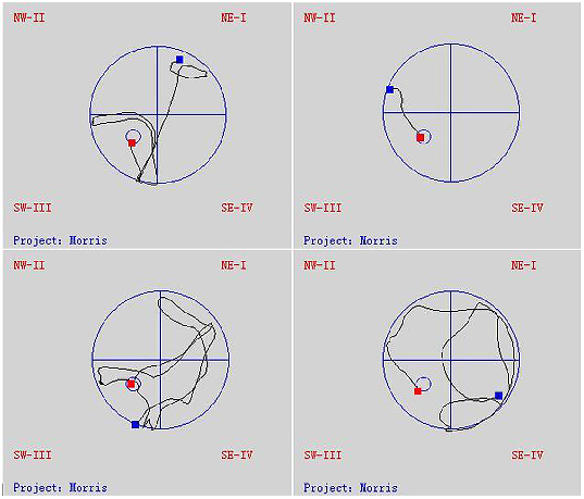
Figure 6
Biochemical markers of oxidative stress and antioxidant capacity in brain tissue are compared across treatment groups, showing dose-dependent protective effects.
chart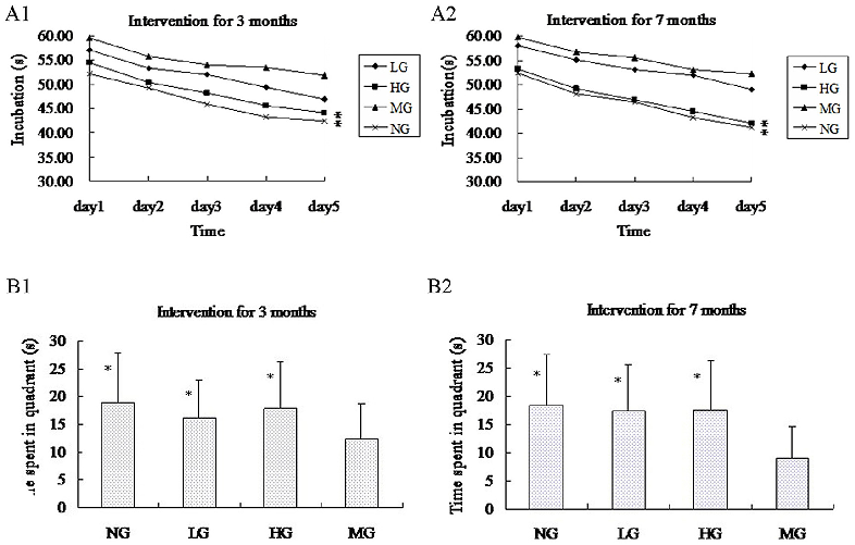
Figure 7
Morris water maze test results reveal that nutrient-supplemented APP-PSN mice demonstrate shorter escape latency and improved spatial memory compared to untreated transgenic controls.
chartFigure 8
MicrographSource Paper
Protective Effects of Dietary Supplementation with a Combination of Nutrients in a Transgenic Mouse Model of Alzheimer's Disease.Cite This Figure
 > Source: Shengyuan Wang et al. "Protective Effects of Dietary Supplementation with a Combination of Nutrients in." *PloS one*, 2015. PMID: [26606074](https://pubmed.ncbi.nlm.nih.gov/26606074/)
<figure>
  <img src="https://pdfs.citedhealth.com/figures/26606074/242.png" alt="Immunofluorescence staining of amyloid-beta plaques in the temporal cortex and hippocampus shows reduced plaque burden in nutrient-treated APP-PSN transgenic mice." />
  <figcaption>Figure 8. Immunofluorescence staining of amyloid-beta plaques in the temporal cortex and hippocampus shows reduced plaque burden in nutrient-treated APP-PSN transgenic mice.<br>  Source: Shengyuan Wang et al. "Protective Effects of Dietary Supplementation with a Combination of Nutrients in." <em>PloS one</em>, 2015. PMID: <a href="https://pubmed.ncbi.nlm.nih.gov/26606074/">26606074</a></figcaption>
</figure>