Descripción
Neuroacanthocytosis encompasses multiple genetic subtypes with overlapping clinical features. This figure provides additional clinical, pathological, or molecular data supporting the differential diagnosis of these rare movement disorders.
More Figures from This Paper
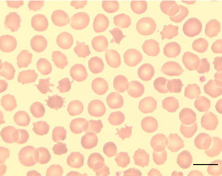
Figure 12
Peripheral blood smear from a patient with McLeod syndrome reveals acanthocytosis, characterized by irregularly spiculated red blood cells. May Gruenwald-Giemsa staining at 100x magnification (scale bar = 10 micrometers) highlights the distinctive thorny morphology of these erythrocytes.
micrograph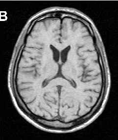
Figure 13
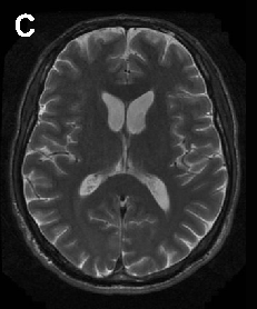
Brain imaging in neuroacanthocytosis typically reveals caudate nucleus atrophy and putaminal changes. This figure presents structural neuroimaging findings characteristic of advanced basal ganglia degeneration in NA patients.
photograph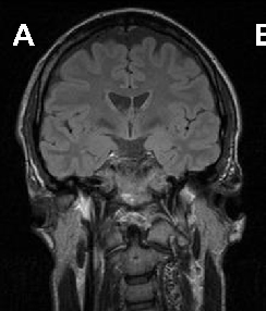
Figure 14
Brain imaging in neuroacanthocytosis typically reveals caudate nucleus atrophy and putaminal changes. This figure presents structural neuroimaging findings characteristic of advanced basal ganglia degeneration in NA patients.
photograph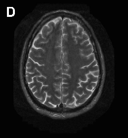
Figure 15
Brain imaging in neuroacanthocytosis typically reveals caudate nucleus atrophy and putaminal changes. This figure presents structural neuroimaging findings characteristic of advanced basal ganglia degeneration in NA patients.
photograph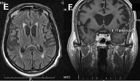
Figure 17
Neuroacanthocytosis encompasses multiple genetic subtypes with overlapping clinical features. This figure provides additional clinical, pathological, or molecular data supporting the differential diagnosis of these rare movement disorders.
diagram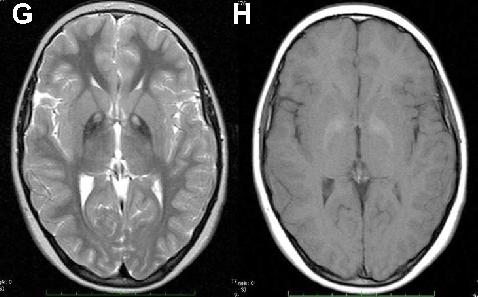
Figure 18
Neuroacanthocytosis encompasses multiple genetic subtypes with overlapping clinical features. This figure provides additional clinical, pathological, or molecular data supporting the differential diagnosis of these rare movement disorders.
diagramCite This Figure
 > Source: Hans H Jung et al. "Neuroacanthocytosis syndromes.." *Orphanet journal of rare diseases*, 2011. PMID: [22027213](https://pubmed.ncbi.nlm.nih.gov/22027213/)
<figure>
  <img src="https://pdfs.citedhealth.com/figures/22027213/139.png" alt="Neuroacanthocytosis encompasses multiple genetic subtypes with overlapping clinical features. This figure provides additional clinical, pathological, or molecular data supporting the differential diagnosis of these rare movement disorders." />
  <figcaption>Figure 16. Neuroacanthocytosis encompasses multiple genetic subtypes with overlapping clinical features. This figure provides additional clinical, pathological, or molecular data supporting the differential diagnosis of these rare movement disorders.<br>  Source: Hans H Jung et al. "Neuroacanthocytosis syndromes.." <em>Orphanet journal of rare diseases</em>, 2011. PMID: <a href="https://pubmed.ncbi.nlm.nih.gov/22027213/">22027213</a></figcaption>
</figure>