विवरण
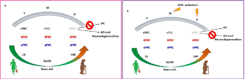
Schematic representation of PKC isoform and RACK protein distribution in membrane fractions of young versus aged rat hippocampus illustrates the age-dependent loss of neuroprotective mechanisms.
More Figures from This Paper
Figure 5
Subcellular distribution of PKC isoforms between cytosolic and membrane fractions is analyzed, showing altered translocation patterns in aged brain tissue.
chart
Figure 6
RACK1 and RACK2 scaffolding protein levels are measured in aging hippocampus, indicating reduced anchoring capacity for activated PKC isoforms.
chart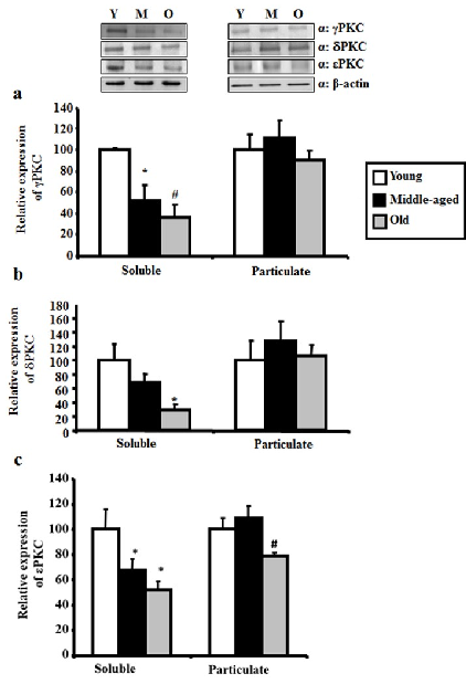
Figure 7
Western blot analysis of gamma, delta, and epsilon PKC levels in hippocampus across young, middle-aged, and aged rats demonstrates progressive age-related reduction.
chart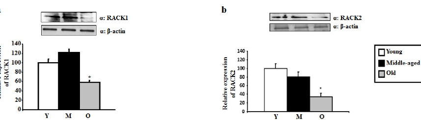
Figure 8
Quantitative densitometry of PKC isoform bands confirms statistically significant declines in neuroprotective PKC levels with advancing age.
chart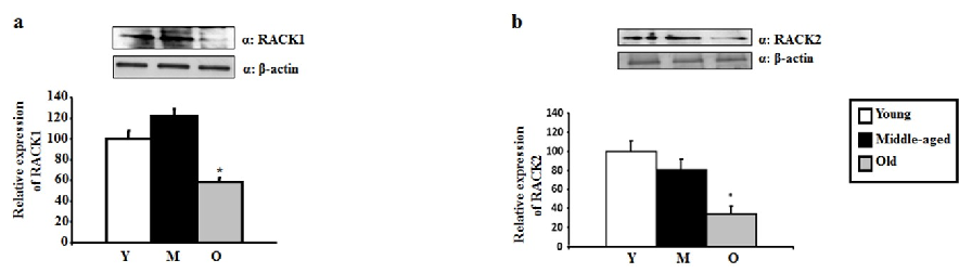
Figure 9
Activated PKC translocation to membrane fractions is diminished in aged rats, with reduced binding to RACK scaffolding proteins contributing to impaired neuroprotective signaling.
chart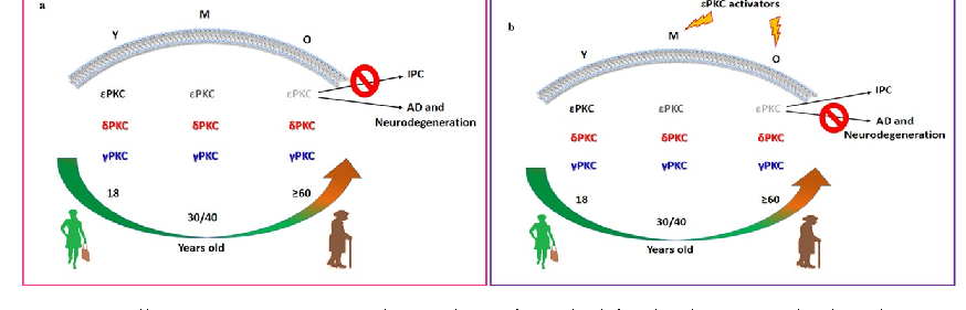
Figure 11
RACK1 and RACK2 protein levels in hippocampal tissue are quantified by Western blot across three age groups, with both scaffolding proteins showing age-related reductions.
chartFigure 10
DiagramSource Paper
Age-Dependent Levels of Protein Kinase Cs in Brain: Reduction of Endogenous Mechanisms of Neuroprotection.Cite This Figure
 > Source: Donatella Pastore et al. "Age-Dependent Levels of Protein Kinase Cs in Brain: Reduction of Endogenous Mech." *International journal of molecular sciences*, 2019. PMID: [31331067](https://pubmed.ncbi.nlm.nih.gov/31331067/)
<figure>
  <img src="https://pdfs.citedhealth.com/figures/31331067/140.png" alt="Schematic representation of PKC isoform and RACK protein distribution in membrane fractions of young versus aged rat hippocampus illustrates the age-dependent loss of neuroprotective mechanisms." />
  <figcaption>Figure 10. Schematic representation of PKC isoform and RACK protein distribution in membrane fractions of young versus aged rat hippocampus illustrates the age-dependent loss of neuroprotective mechanisms.<br>  Source: Donatella Pastore et al. "Age-Dependent Levels of Protein Kinase Cs in Brain: Reduction of Endogenous Mech." <em>International journal of molecular sciences</em>, 2019. PMID: <a href="https://pubmed.ncbi.nlm.nih.gov/31331067/">31331067</a></figcaption>
</figure>