Vitamin E चित्र
1 सहकर्मी-समीक्षित शोध से आंकड़े
सभी
Omega-3 Fatty Acids (DHA/EPA)
Bacopa monnieri
Citicoline
Phosphatidylserine
Ginkgo biloba
Curcumin
Creatine
Rhodiola rosea
L-Theanine
Panax Ginseng
Vitamin D
Vitamin B12
Folate
Resveratrol
Alpha-Lipoic Acid
Vitamin E
Green Tea Extract (EGCG)
Zinc
Uridine Monophosphate
Taurine
Lutein & Zeaxanthin
Melatonin
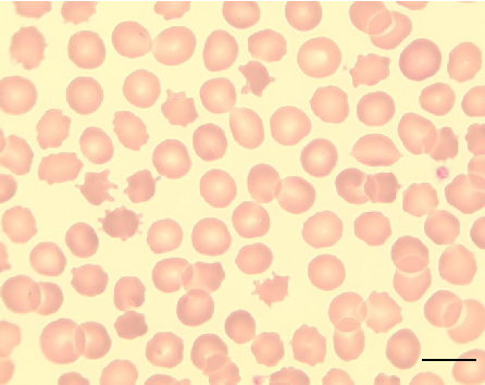
Figure 12
Micrograph
Peripheral blood smear from a patient with McLeod syndrome reveals acanthocytosis, characterized by irregularly spiculated red blood cells. May Gruenwald-Giemsa staining at 100x magnification (scale bar = 10 micrometers) highlights the distinctive thorny morphology of these erythrocytes.
Neuroacanthocytosis syndromes.