Quy trình nghiên cứu
368 hình ảnh từ nghiên cứu có bình duyệt
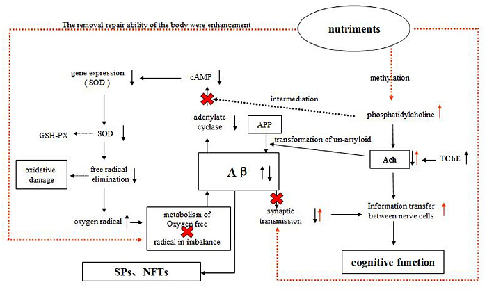
A schematic diagram illustrates the proposed mechanisms by which the compound nutrient mixture may protect against Alzheimer's pathology, including acetylcholine and cAMP signaling.
Protective Effects of Dietary Supplementation with a Combination of Nutrients in a …
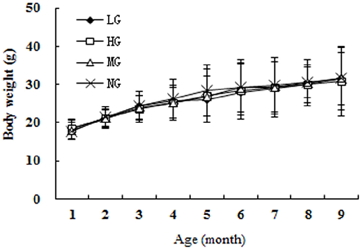
Daily food intake across groups confirms consistent consumption, with no significant differences between the low, high, and model groups during the study.
Protective Effects of Dietary Supplementation with a Combination of Nutrients in a …
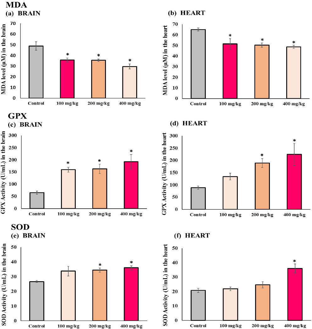
Biochemical markers of oxidative stress and antioxidant capacity in brain tissue are compared across treatment groups, showing dose-dependent protective effects.
Protective Effects of Dietary Supplementation with a Combination of Nutrients in a …
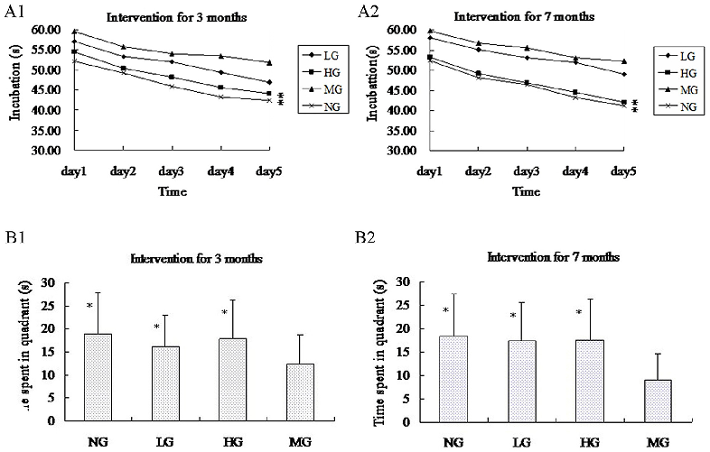
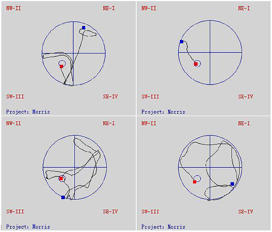
Morris water maze test results reveal that nutrient-supplemented APP-PSN mice demonstrate shorter escape latency and improved spatial memory compared to untreated transgenic controls.
Protective Effects of Dietary Supplementation with a Combination of Nutrients in a …
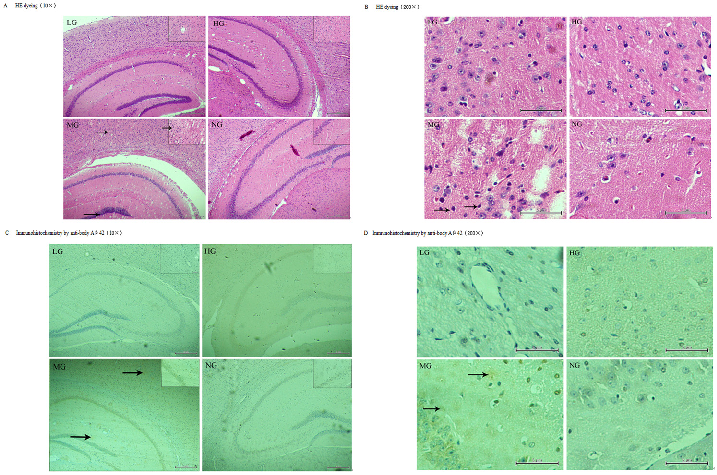
Immunofluorescence staining of amyloid-beta plaques in the temporal cortex and hippocampus shows reduced plaque burden in nutrient-treated APP-PSN transgenic mice.
Protective Effects of Dietary Supplementation with a Combination of Nutrients in a …
Subcellular distribution of PKC isoforms between cytosolic and membrane fractions is analyzed, showing altered translocation patterns in aged brain tissue.
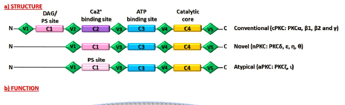
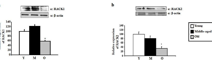
Age-Dependent Levels of Protein Kinase Cs in Brain: Reduction of Endogenous Mechanisms …
RACK1 and RACK2 scaffolding protein levels are measured in aging hippocampus, indicating reduced anchoring capacity for activated PKC isoforms.
Age-Dependent Levels of Protein Kinase Cs in Brain: Reduction of Endogenous Mechanisms …
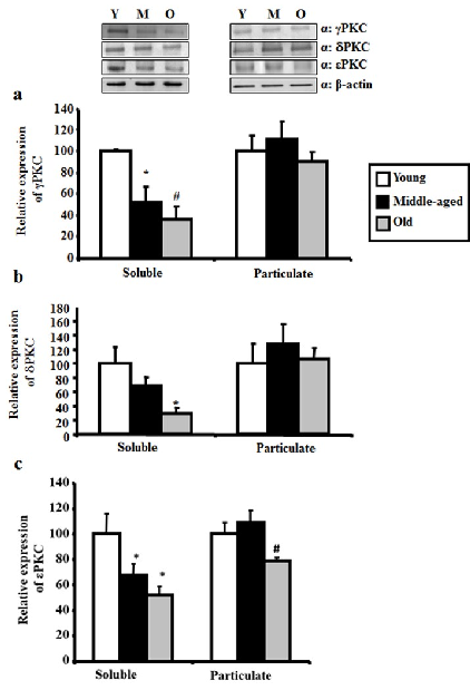
Western blot analysis of gamma, delta, and epsilon PKC levels in hippocampus across young, middle-aged, and aged rats demonstrates progressive age-related reduction.
Age-Dependent Levels of Protein Kinase Cs in Brain: Reduction of Endogenous Mechanisms …
Quantitative densitometry of PKC isoform bands confirms statistically significant declines in neuroprotective PKC levels with advancing age.
Age-Dependent Levels of Protein Kinase Cs in Brain: Reduction of Endogenous Mechanisms …
Activated PKC translocation to membrane fractions is diminished in aged rats, with reduced binding to RACK scaffolding proteins contributing to impaired neuroprotective signaling.
Age-Dependent Levels of Protein Kinase Cs in Brain: Reduction of Endogenous Mechanisms …
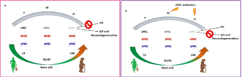
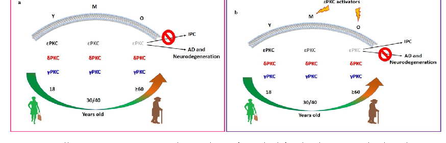
Schematic representation of PKC isoform and RACK protein distribution in membrane fractions of young versus aged rat hippocampus illustrates the age-dependent loss of neuroprotective mechanisms.
Age-Dependent Levels of Protein Kinase Cs in Brain: Reduction of Endogenous Mechanisms …
RACK1 and RACK2 protein levels in hippocampal tissue are quantified by Western blot across three age groups, with both scaffolding proteins showing age-related reductions.
Age-Dependent Levels of Protein Kinase Cs in Brain: Reduction of Endogenous Mechanisms …
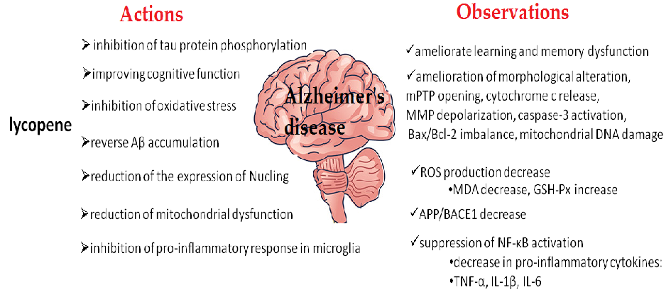
Introductory overview of the carotenoid compounds evaluated for neuroprotective effects in Alzheimer's disease. The review examines how these dietary pigments may counteract neurodegeneration through antioxidant and anti-inflammatory pathways.
Carotenoid Supplementation for Alleviating the Symptoms of Alzheimer's Disease.
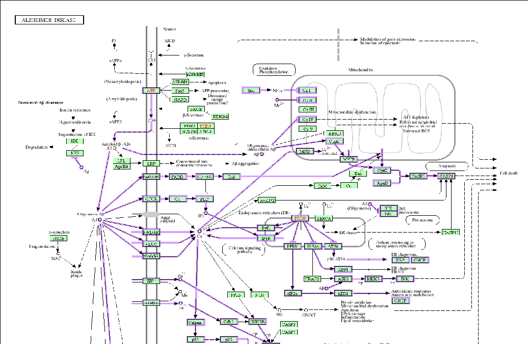
![Figure 1. Alzheimer’s disease KEGG pathway (hsa05010; Alzheimer disease—Homo sapiens (human)) generated online at https://www.genome.jp/kegg-bin/show_pathway?hsa05010, accessed on 8 March 2024) [115].](https://pdfs.citedhealth.com/figures/39201668/171.png)
KEGG pathway map (hsa05010) illustrating the molecular cascade involved in Alzheimer's disease pathogenesis in humans. The diagram highlights potential intervention points where carotenoid compounds may modulate amyloid-beta accumulation, tau phosphorylation, and neuroinflammatory signaling.
Carotenoid Supplementation for Alleviating the Symptoms of Alzheimer's Disease.
Evidence summary on the association between specific carotenoid compounds and Alzheimer's disease biomarkers. The data suggest that higher carotenoid status may be linked to reduced oxidative damage in neural tissues.
Carotenoid Supplementation for Alleviating the Symptoms of Alzheimer's Disease.
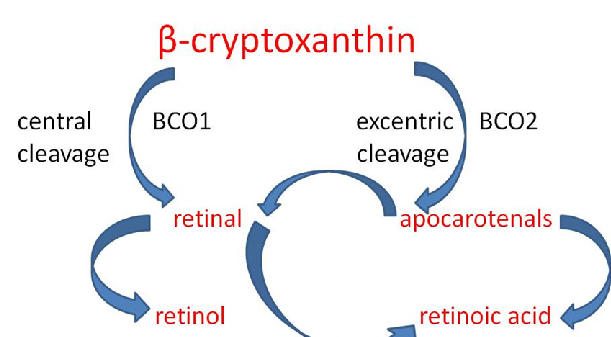
Alpha- and beta-cryptoxanthin serve as provitamin A sources due to their unsubstituted beta-ring structure. The review discusses their potential role in Alzheimer's disease prevention through both antioxidant activity and vitamin A metabolite production in neural tissue.
Carotenoid Supplementation for Alleviating the Symptoms of Alzheimer's Disease.
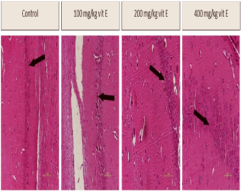
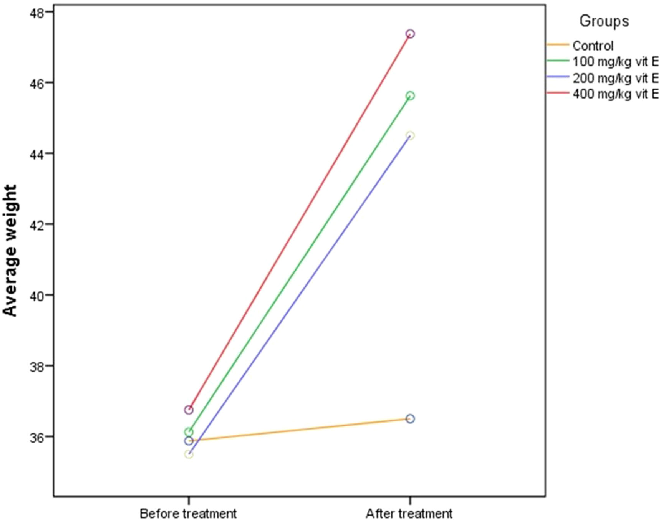
Oxidative stress biomarker measurements across vitamin E dosage groups in aged mice. The results indicate a dose-dependent reduction in oxidative damage markers, suggesting vitamin E supplementation may attenuate age-related oxidative stress.
The Impact of Vitamin E Supplementation on Oxidative Stress, Cognitive Functions, and …
Cognitive function assessment results comparing aged mice receiving different vitamin E doses. Behavioral testing data suggest that vitamin E supplementation is associated with improved cognitive performance in a dose-dependent manner.
The Impact of Vitamin E Supplementation on Oxidative Stress, Cognitive Functions, and …
Gene expression analysis in aged mice treated with varying vitamin E doses, examining aging-related molecular pathways. The data reveal differential regulation of oxidative stress response genes across treatment groups.
The Impact of Vitamin E Supplementation on Oxidative Stress, Cognitive Functions, and …
Additional molecular or behavioral endpoint data from the vitamin E dose-response study in aged mice. The comprehensive assessment supports a beneficial role for vitamin E in mitigating multiple aging-related phenotypes.
The Impact of Vitamin E Supplementation on Oxidative Stress, Cognitive Functions, and …
![Following oral administration, curcumin demonstrates poor systemic bioavailability due to limited intestinal absorption, rapid hepatic metabolism, and swift systemic elimination [23]. Clinical pharmacokinetic studies have shown that even high oral doses (](https://pdfs.citedhealth.com/figures/40944272/199.png)
Overview of curcumin's pharmacokinetic challenges following oral administration, including poor systemic bioavailability due to limited intestinal absorption and rapid hepatic metabolism. The narrative review discusses strategies to enhance curcumin delivery to the central nervous system for neuroprotective applications.
The Neuroprotective Role of Curcumin: From Molecular Pathways to Clinical Translation-A Narrative …
![Curcumin exerts broad-spectrum anti-inflammatory effects predominantly via modulation of the nuclear factor kappa B (NF-κB) signaling pathway [70]. By inhibiting NF-κB activation, curcumin suppresses the transcription of pivotal pro-inflammatory mediators](https://pdfs.citedhealth.com/figures/40944272/226.png)
Curcumin's anti-inflammatory mechanisms operate primarily through modulation of the NF-kappa-B signaling pathway. The diagram illustrates how curcumin suppresses neuroinflammatory cascades implicated in Alzheimer's disease, Parkinson's disease, and post-stroke cognitive impairment.
The Neuroprotective Role of Curcumin: From Molecular Pathways to Clinical Translation-A Narrative …
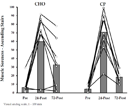
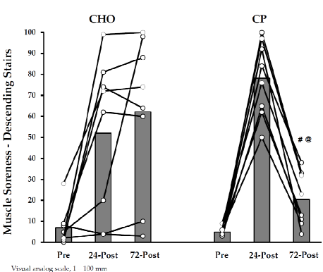
Mid-study recovery data comparing protein-supplemented and control groups following marathon running. The time-course analysis captures the progression of muscle recovery indicators.
Protein Supplementation During or Following a Marathon Run Influences Post-Exercise Recovery.
Extended recovery assessment data for marathon runners, comparing outcomes between those who received protein supplementation during or after the race and those who did not.
Protein Supplementation During or Following a Marathon Run Influences Post-Exercise Recovery.
Trang 11 / 16