अनुसंधान प्रक्रिया
87 सहकर्मी-समीक्षित शोध से आंकड़े
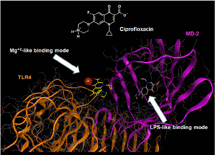
Molecular docking analysis reveals two alternative binding conformations of ciprofloxacin within the TLR4-MD-2 complex binding pocket, suggesting direct physical interaction with the innate immune receptor.
Ciprofloxacin and levofloxacin attenuate microglia inflammatory response via TLR4/NF-kB pathway.
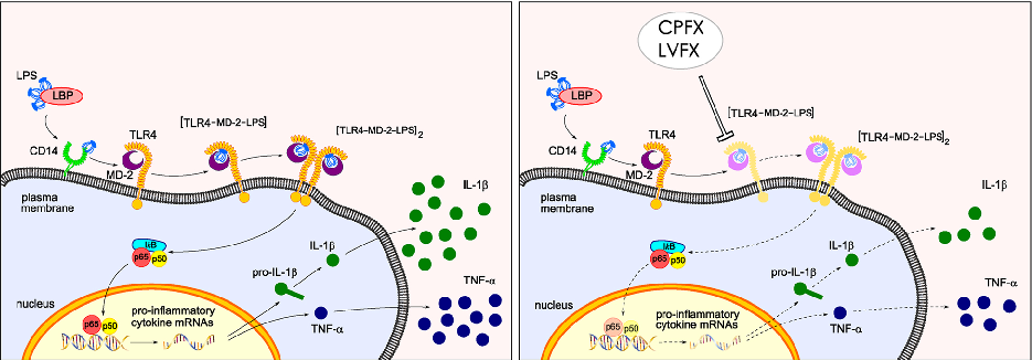
Proposed mechanistic model depicts how ciprofloxacin and levofloxacin target the TLR4-MD-2 complex to block LPS-induced downstream signaling cascades and cytokine production.
Ciprofloxacin and levofloxacin attenuate microglia inflammatory response via TLR4/NF-kB pathway.
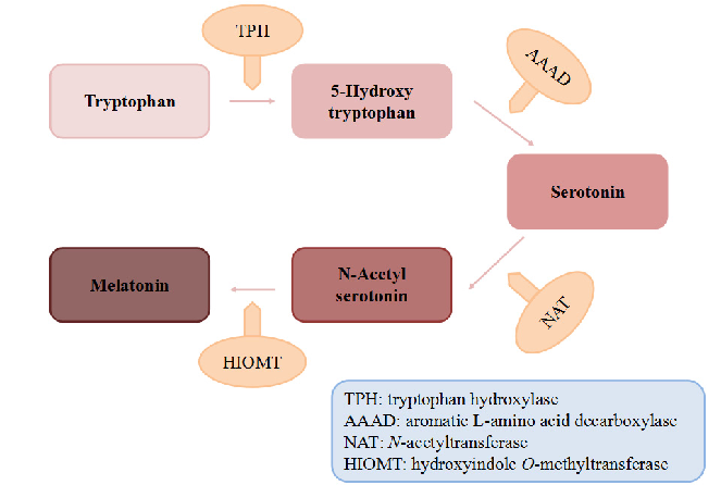
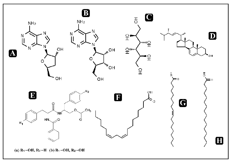
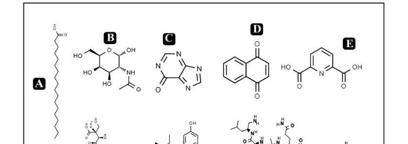
Biosynthetic pathway of melatonin from tryptophan is displayed, showing the sequential enzymatic steps through serotonin N-acetyltransferase and hydroxyindole-O-methyltransferase.
Dietary Sources and Bioactivities of Melatonin.
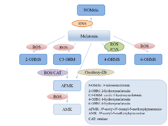
Melatonin and its metabolites including 6-hydroxymelatonin, AFMK, and AMK are structurally depicted, illustrating the biotransformation cascade.
Dietary Sources and Bioactivities of Melatonin.
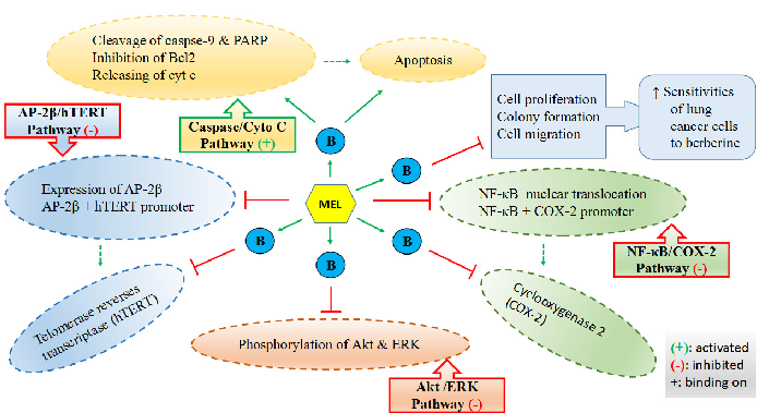
Mechanisms by which melatonin enhances lung cancer cell sensitivity to berberine are diagrammed, showing synergistic effects on apoptotic and autophagic pathways.
Dietary Sources and Bioactivities of Melatonin.
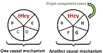
A causal model illustrates how elevated plasma homocysteine may contribute to dementia through multiple pathways, interacting with other risk factors such as age, hypercholesterolemia, and genetic predisposition. No single factor is sufficient alone; rather, combinations of component causes drive disease.
Homocysteine and Dementia: An International Consensus Statement.
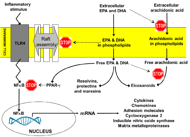
Key anti-inflammatory actions of EPA and DHA include suppression of NF-kB activation, reduction of pro-inflammatory eicosanoid production, inhibition of NLRP3 inflammasome activation, and promotion of regulatory T-cell differentiation through PPAR-gamma signaling.
Expert Opinion on Benefits of Long-Chain Omega-3 Fatty Acids (DHA and EPA) …
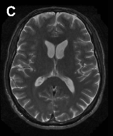
Neuroacanthocytosis encompasses multiple genetic subtypes with overlapping clinical features. This figure provides additional clinical, pathological, or molecular data supporting the differential diagnosis of these rare movement disorders.
Neuroacanthocytosis syndromes.
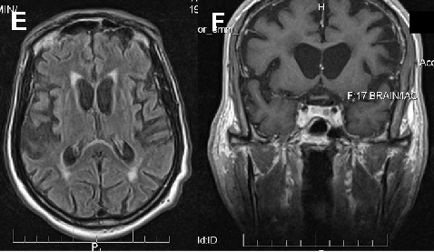
Neuroacanthocytosis encompasses multiple genetic subtypes with overlapping clinical features. This figure provides additional clinical, pathological, or molecular data supporting the differential diagnosis of these rare movement disorders.
Neuroacanthocytosis syndromes.
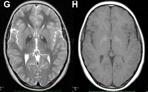
Neuroacanthocytosis encompasses multiple genetic subtypes with overlapping clinical features. This figure provides additional clinical, pathological, or molecular data supporting the differential diagnosis of these rare movement disorders.
Neuroacanthocytosis syndromes.
Neuroprotective and anti-fatigue properties of cordycepin have been demonstrated in preclinical models. This figure highlights cordycepin's potential benefits for neurological health and physical performance.
Cordycepin for Health and Wellbeing: A Potent Bioactive Metabolite of an Entomopathogenic …
Cordycepin modulates immune responses through effects on macrophage activation and cytokine production. This figure presents immunomodulatory data from in vitro and in vivo studies of cordycepin treatment.
Cordycepin for Health and Wellbeing: A Potent Bioactive Metabolite of an Entomopathogenic …
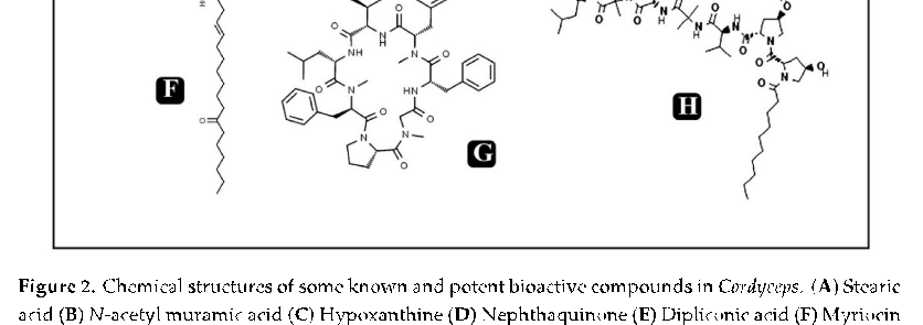
Anti-microbial and anti-viral activities of cordycepin complement its anti-inflammatory properties. This figure summarizes evidence for cordycepin's broad-spectrum antimicrobial potential.
Cordycepin for Health and Wellbeing: A Potent Bioactive Metabolite of an Entomopathogenic …

Preclinical evidence supports cordycepin's hepatoprotective and nephroprotective properties. This figure presents data on cordycepin's organ-protective effects in various disease models.
Cordycepin for Health and Wellbeing: A Potent Bioactive Metabolite of an Entomopathogenic …
![Figure 4. (A) Possible mechanism of cordycepin for its anti-diabetic activity (B) Possible mechanism of cordycepin in regulation of fat metabolism in hyperlipidemia [47].](https://pdfs.citedhealth.com/figures/32545666/197.png)
Cordycepin may exert anti-diabetic effects through regulation of glucose metabolism and insulin sensitivity. Panel A illustrates the proposed mechanism for anti-diabetic activity, while Panel B depicts cordycepin's role in fat metabolism regulation in hyperlipidemia.
Cordycepin for Health and Wellbeing: A Potent Bioactive Metabolite of an Entomopathogenic …
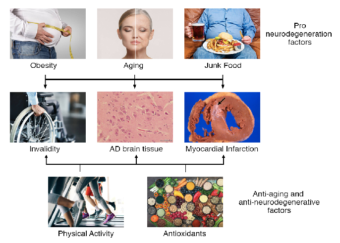
Pro-degeneration factors including aging, obesity, and unhealthy diets can be counterbalanced by physical activity, caloric restriction, and antioxidants. This diagram illustrates how these opposing influences modulate the onset, severity, and duration of neurodegenerative diseases including Alzheimer's, Parkinson's, and ALS.
Dietary habits, lifestyle factors and neurodegenerative diseases.
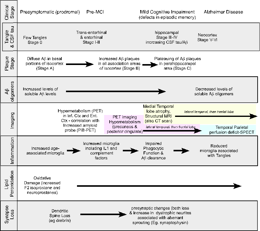
Alzheimer's disease involves a complex pathological cascade initially triggered by amyloid-beta accumulation or aberrant APP processing. This figure argues for pleiotropic interventions that simultaneously target multiple pathological mechanisms rather than single molecular targets.
Why pleiotropic interventions are needed for Alzheimer's disease.
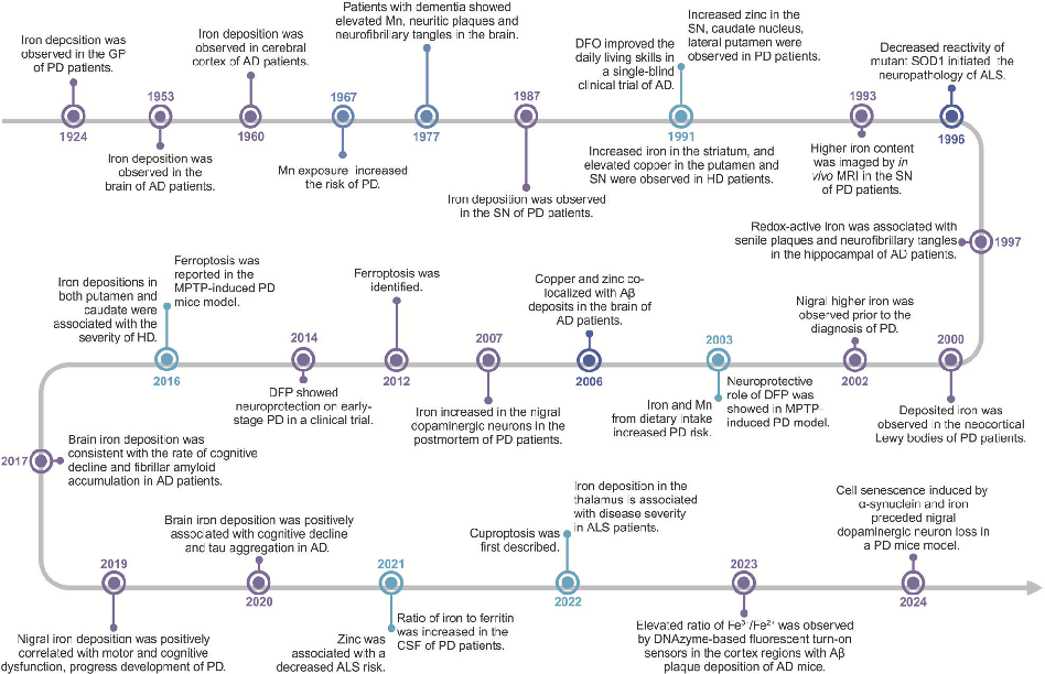
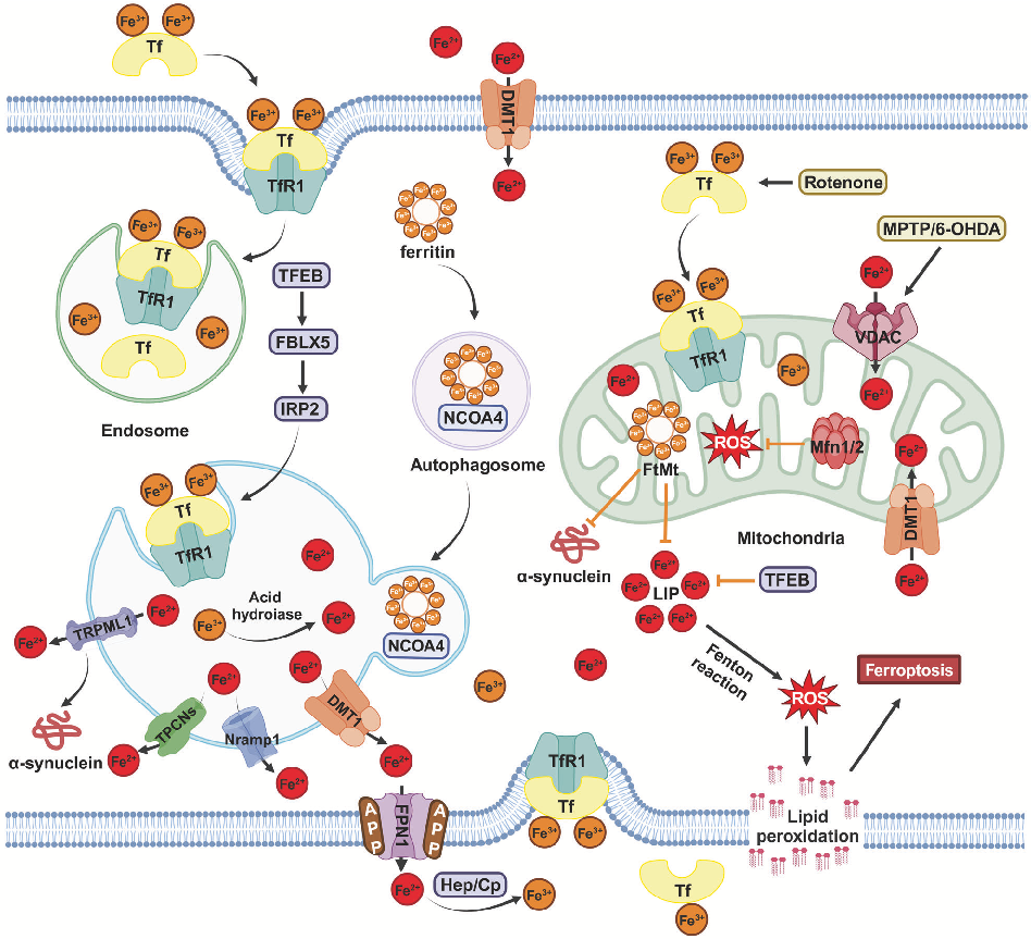
Molecular pathways of iron metabolism and ferroptosis in the context of neurodegenerative disease. Excessive iron accumulation in neurons can trigger lipid peroxidation and cell death through ferroptotic mechanisms.
Homeostasis and metabolism of iron and other metal ions in neurodegenerative diseases.
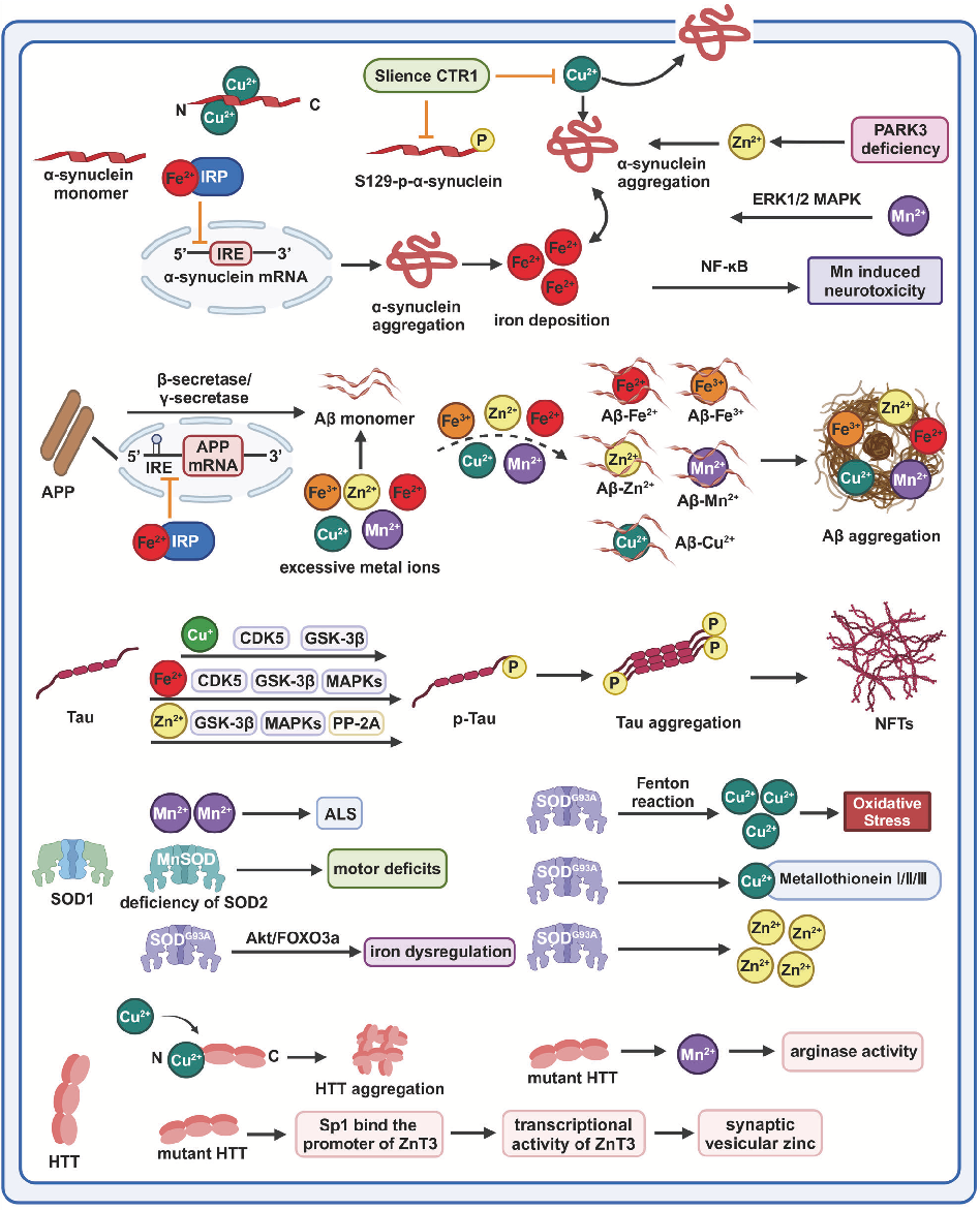
Schematic of copper homeostasis and cuproptosis mechanisms relevant to neurodegeneration. Copper imbalance has been implicated in the pathogenesis of Wilson's disease and may contribute to Alzheimer's disease progression.
Homeostasis and metabolism of iron and other metal ions in neurodegenerative diseases.
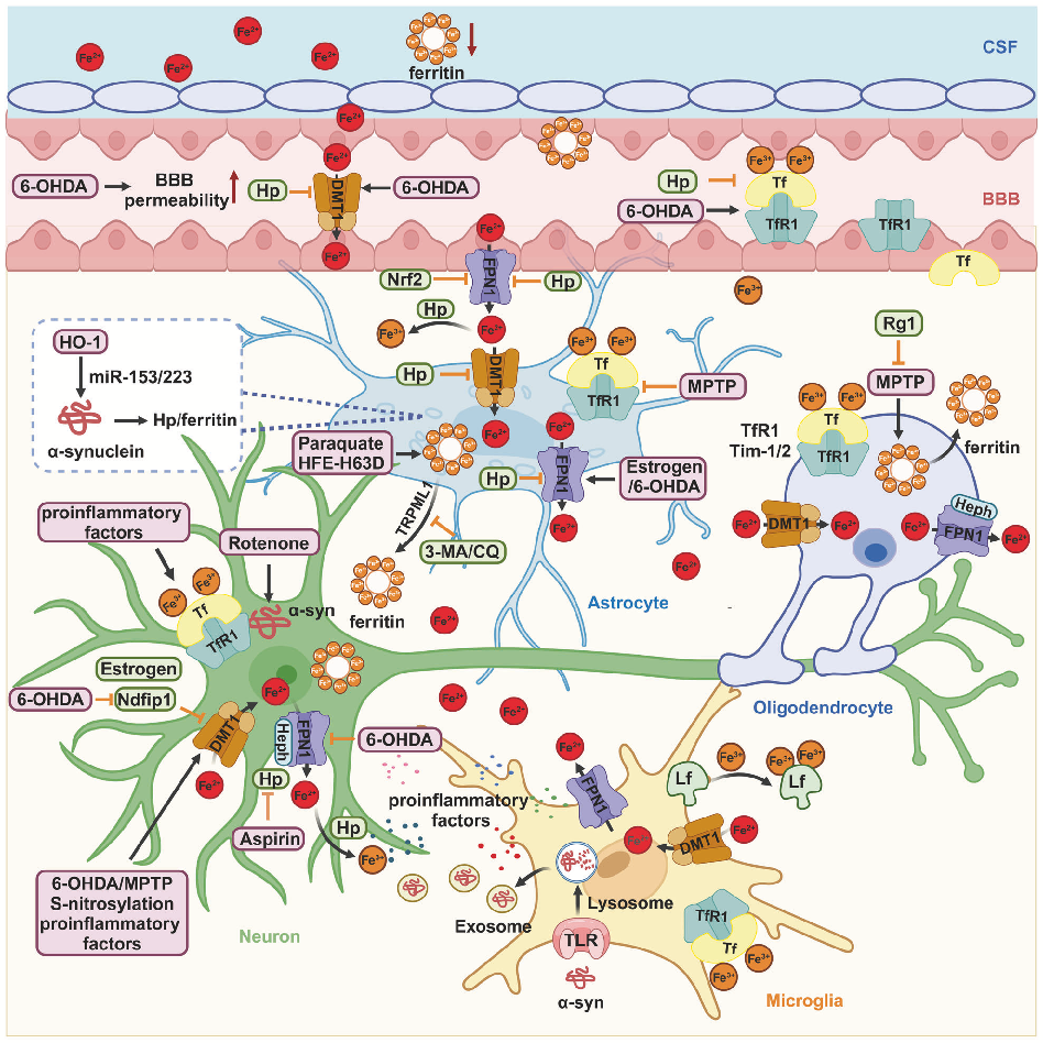
Illustration of zinc and manganese transport and regulatory mechanisms in the central nervous system, highlighting how disruptions in metal ion balance may accelerate neurodegenerative processes.
Homeostasis and metabolism of iron and other metal ions in neurodegenerative diseases.
Summary of therapeutic strategies targeting metal ion dysregulation in neurodegenerative diseases, including iron chelation, antioxidant supplementation, and metal transporter modulation.
Homeostasis and metabolism of iron and other metal ions in neurodegenerative diseases.
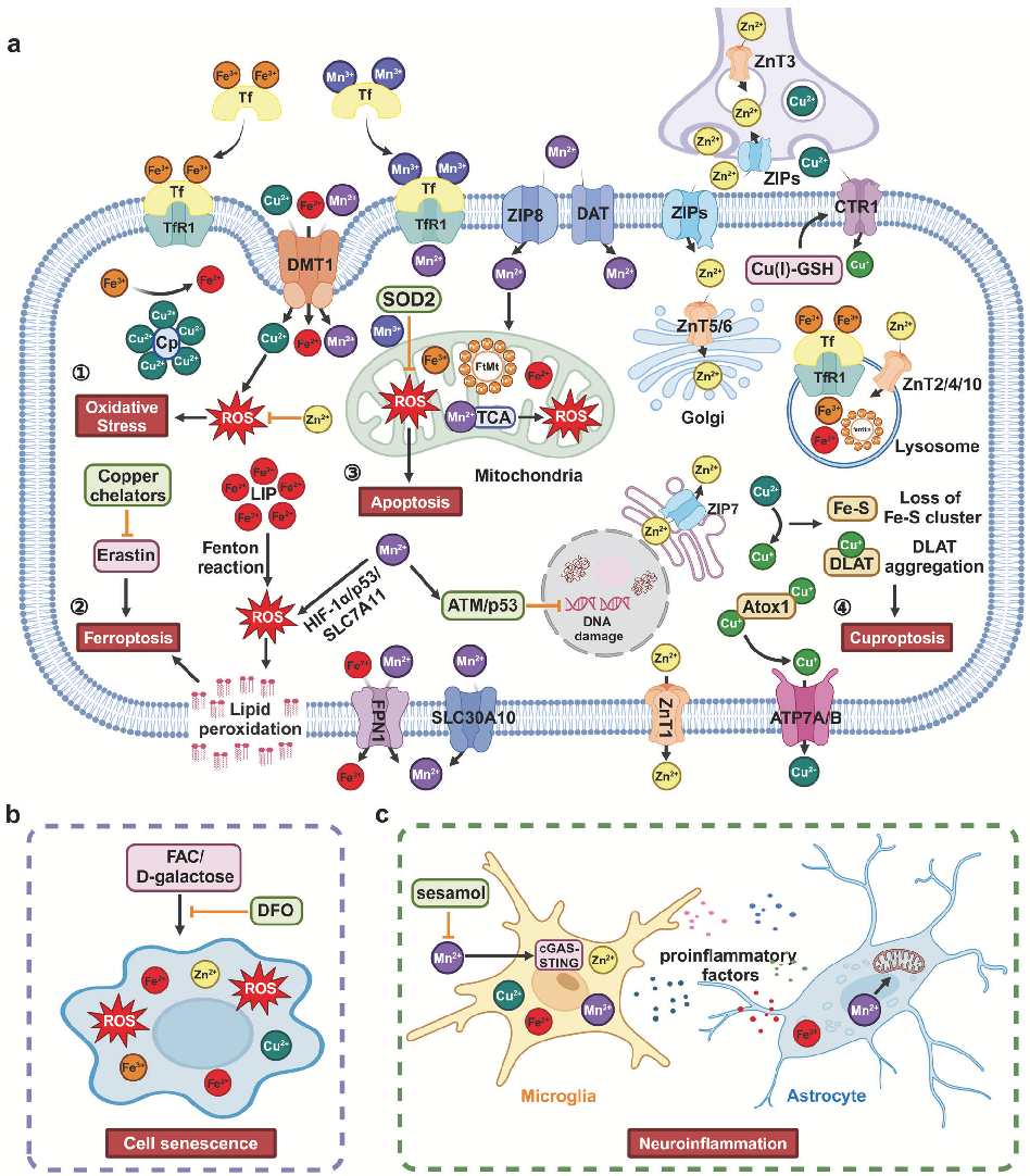
Integrative model linking metal ion imbalance to multiple neurodegenerative disease mechanisms including oxidative stress, ferroptosis, cuproptosis, cellular senescence, and neuroinflammation.
Homeostasis and metabolism of iron and other metal ions in neurodegenerative diseases.
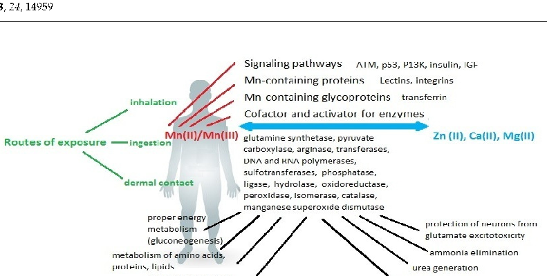
Neurological consequences of chronic manganese exposure are summarized, including cognitive impairment, motor dysfunction, and emotional disturbances. The symptoms collectively termed manganism share clinical features with Parkinson's disease.
Consequences of Disturbing Manganese Homeostasis.
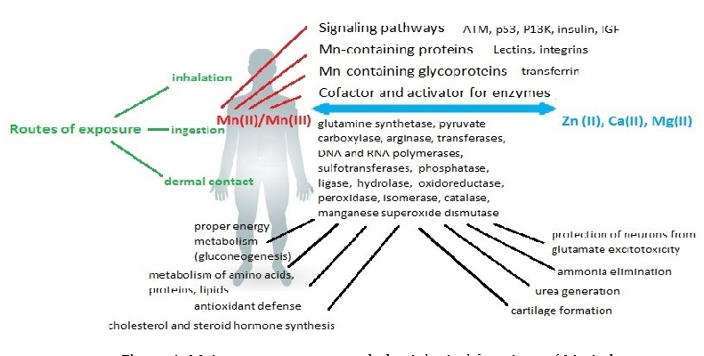
Manganese's dual role as both essential nutrient and potential toxin is explored through its effects on enzymatic systems. Superoxide dismutase, arginase, and glutamine synthetase all require manganese as a cofactor for normal function.
Consequences of Disturbing Manganese Homeostasis.
पृष्ठ 1 / 4